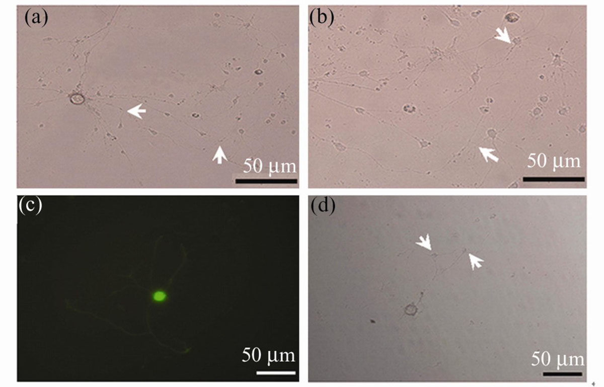
Fig. 3
Characterization of major pelvic ganglion neurons and spinal motor neurons in co-culture
Download original image
(a), (b): Phase contrast images of a major pelvic ganglion neuron contacted by multiple spinal motor neurons. (c), (d): GFP-labelled major pelvic ganglion neurons and spinal motor neurons were shown in proximity to each other. Some apparent SMN-MPG neuron contacts are seen in (a), (b) and (d) (arrows)
Current usage metrics show cumulative count of Article Views (full-text article views including HTML views, PDF and ePub downloads, according to the available data) and Abstracts Views on Vision4Press platform.
Data correspond to usage on the plateform after 2015. The current usage metrics is available 48-96 hours after online publication and is updated daily on week days.
Initial download of the metrics may take a while.