| Issue |
Wuhan Univ. J. Nat. Sci.
Volume 29, Number 5, October 2024
|
|
|---|---|---|
| Page(s) | 461 - 470 | |
| DOI | https://doi.org/10.1051/wujns/2024295461 | |
| Published online | 20 November 2024 | |
Biology
CLC number: O614
Synthesis, Cytotoxicity, Apoptosis and Cell Cycle Arrest of a Ruthenium(II)-Substituted Keggin Polyoxotungstate
钌多取代Keggin-型多钨酸盐的合成、细胞毒性、细胞凋亡和细胞周期研究
College of Chemical Engineering and Technology, Taiyuan University of Science and Technology, Taiyuan 030024, Shanxi, China
† Corresponding author. E-mail: This email address is being protected from spambots. You need JavaScript enabled to view it.
Received:
15
September
2023
Abstract
The ruthenium multi-substituted polyoxotungstate, K7[SiW9O37Ru4(H2O)3Cl3]·15H2O (S1), was synthesized by a conventional aqueous solution containing the trilacunary Keggin-anions β-Na9HSiW9O34·12H2O (S2) and RuCl3·nH2O (S3). Compound S1 was characterized by elemental analysis, energy-dispersive X-ray spectroscopy (EDS), thermogravimetric analysis (TG), infrared spectroscopy (IR), uliraviolet visible absorption spectroscopy (UV/Vis) and X-ray photoelectron spectroscopy (XPS). The cytotoxicitycy of S1 was tested in C33A (human cervical cancer), DLD-1 (human colon cancer), HepG2 (human liver cancer) and human normal embryonic lung fibroblasts cell (MRC-5). And the viability of these treated cells was evaluated by MTT (3-(4,5-dimethylthiazol-2-yl)-2,5-diphenyltetrazolium bromide) assay.To explore the mode of cell death induced by S1, morphological study of DNA damage and apoptosis assays were conducted. These analyses revealed that S1 exerted its cytotoxic effect in a dose-dependent manner, primarily triggering apoptotic cell death. Cell cycle analysis by flow cytometry indicated that compound S1 caused cell cycle arrest and accumulated cells in S phase.
摘要
本文以三缺位Keggin-型阴离子β-Na9HSiW9O34·12H2O (S2)和RuCl3·nH2O (S3)为原料, 在水溶液中合成了一个钌多取代多钨酸盐, 其化学式为: K7[SiW9O37Ru4(H2O)3Cl3]·15H2O (S1)。通过元素分析、能谱、热重、红外、紫外和X-射线光电子能谱对化合物S1进行了表征。通过MTT法测试了化合物S1对C33A、DLD-1、HepG2三种肿瘤细胞和人正常胚肺成纤维细胞MRC-5的细胞毒性。通过观察细胞形态及流式细胞仪考察了肿瘤细胞的死亡方式。实验结果表明化合物S1诱导肿瘤细胞凋亡而非坏死, 并且细胞存活率与S1的浓度呈梯度关系。最后通过流式细胞仪分析细胞周期的变化, 结果显示化合物S1使细胞周期停留在S期。
Key words: ruthenium multi-substituted polyoxometalate / cytotoxicity / cell apoptosis / cell cycle arrest
关键字 : 钌多取代的多钨酸盐 / 细胞毒性 / 细胞凋亡 / 细胞周期
Cite this article: JIA Shifang, HAO Xiuli, WEN Yanzhen, et al. Synthesis, Cytotoxicity, Apoptosis and Cell Cycle Arrest of a Ruthenium(II)-Substituted Keggin Polyoxotungstate[J]. Wuhan Univ J of Nat Sci, 2024, 29(5): 461-470.
Biography: JIA Shifang, female, Ph.D., research direction: bioinorganic chemistry. E-mail: This email address is being protected from spambots. You need JavaScript enabled to view it.
Fundation item: Supported by the National Natural Science Foundation of China (21701120), the Science and Technology Innovation Project of Colleges and Universities in Shanxi Province (2020L0334), and the Innovation and Entrepreneurship Training Program for College Students in Shanxi Province(20240778)
© Wuhan University 2024
 This is an Open Access article distributed under the terms of the Creative Commons Attribution License (https://creativecommons.org/licenses/by/4.0), which permits unrestricted use, distribution, and reproduction in any medium, provided the original work is properly cited.
0 Introduction
Cancer is a serious public health problem, which has threatened human health on a worldwide scale. At present, chemotherapy has been an important strategy and approach for cancer treatment. Therefore, identifying an effective and low-toxicity drug has become the priority in tumor treatment research[1-3]. In both clinical application and experimental exploration, most chemotherapeutic drugs mainly composed of synthetical compounds and adjuvant chemotherapeutic of natural substances or photochemicals. The synthetical compounds have attracted wide attention due to many advantages, such as designability, controllability and predictability. In recent years, it is worth noting that polyoxometalates (POMs) have become biomedical agents because of their versatile bioactivity including antibacterial, antitumor and antiviral functions[4-7]. Compared with current drugs, POMs hold unique advantages. Their composition, molecular structure and physiochemical properties are adjustable and can be easily synthesized from precursors[8-11]. Moreover, POMs have other advantages. Transition metals can be introduced into their structure to obtain transition metal-substituted POMs, such as mono, dis or tri-substituted polyanions[12-14]. The integration of transition metals with vacancy POMs modifies the original saturated POMs'composition, charge and physicochemical properties, thereby enhancing their biological activity[15,16].
In addition, owing to the low energy barrier between their oxidation states, low atomic radius (0.125 nm) in transition metal series, and high solubility in water, the ruthenium compounds are conducive to accumulation in cancer tissues. Thus, ruthenium compounds have potential value in the development of chemotherapeutic drugs[17,18]. The synergistic interaction between ruthenium and vacancy POMs is expected to endow ruthenium-substituted polyoxometalates (Ru-POMs) with high antitumor activity. Although a multitude of Ru-POMs have been reported[19-27], the antitumor activities of these compounds have rarely been explored or reported. In previous research, we investigated the antitumor activities of a monoruthenium(II)-substituted Dawson polyoxotungstate[28]. However, we noticed that most syntheses of Ru-POMs have predominantly relied on utilizing the monolacunary Keggin [XW11O39]n- (X = P, Ge or Si) or Dawson-type [P2W17O61]10- polyoxoanions as reagents or ligands for coordination with ruthenium[19-25]. And the compounds synthesized using other types of lacunary POMs combined with ruthenium were rarely reported.
Based on the aforementioned considerations, we chose trilacunary POMs β-[SiW9O34]10- (β-SiW9) and RuCl3·nH2O as raw materials to synthesize a ruthenium multi-substituted polyoxometalate compound, designated as K7[SiW9O37Ru4(H2O)3Cl3]·15H2O (S1). The cytotoxicity of compound S1in vitro was evaluated by MTT (3-(4,5-dimethylthiazol-2-yl)-2,5-diphenyltetrazolium bromide) assay. And, the percentage of necrotic and apoptotic C33A (human cervical cancer) cells induced by the complex were also investigated by fluorescence microscopy and flow cytometry. Furthermore, the cell cycle distribution within C33A cells was detected by flow cytometry. Our results indicate that compound S1 can inhibit cell proliferation and induce apoptosis in cancer cells.
1 Experimental
1.1 Materials and Instrumentation
All reagents and solvents were commercially sourced unless otherwise specified. Ultrapure Milli-Q water was used in all experiments. β-Na9HSiW9O34·12H2O was prepared according to the reported methods[29]. RPMI 1640 was purchased from Sigma. C33A, DLD-1 (human colon cancer), HepG2 (human liver cancer), MRC-5 (human normal embryonic lung fibroblasts) cell lines were purchased from the American Type Culture Collection (Rockville, MD, USA). The elemental analysis for W, Ru, and K was performed using a Leeman inductively coupled plasma (ICP) spectrometer. Cl was analyzed by ion chromatography. Energy-dispersive X-ray spectroscopy (EDS) was performed by X-Max Extreme. Thermogravimetric analysis (TG) was carried out on a Pyris Diamond TG instrument under N2 with a heating rate of 10 ℃·min-1. Infrared spectra were recorded on a Perkin-Elmer Spectrum FT-IR (fourier-transform infrared spectroscopy) Spectrometer using KBr pellets. Raman spectrum was tested by the Raman spectrometer. Electronic absorption spectra (UV/Vis) were recorded on a Lambda 750 spectrophotometer. X-ray photoelectron spectroscopy (XPS) of Ru and W were collected on a VG ESCALAB MK II spectrometer equipped with a Mg Kα (1 253.6 eV) achromatic X-ray source. Solution electronic emission spectra in acetonitrile was recorded by Shimadzu 3100 spectrophotometer and Shimadzu RF-5301 PC spectrofluorometer. Cyclic voltammetry (CV) was obtained with CHI 630E instrument in a three-electrode system: a glassy carbon electrode (GCE, diameter 3 mm) served as the working electrode, a platinum wire acted as the counter electrode, and Ag/AgCl was the reference electrode.
1.2 Synthesis
The synthesis of S1 was performed as follows:
About 0.27 g of RuCl3·nH2O (corresponding to 1.1 mmol) was dissolved in 8 mL of water. Approximately one hour later, 1.0 g of solid β-Na9HSiW9O34·12H2O (equivalent to 0.37 mmol) was slowly added with vigorous stirring. The pH of the solution was subsequently adjusted to 5.5 using aqueous Na2CO3 (1 mol/L). Then the solution was heated to a temperature range of 70 - 80 ℃ and was stirred for half an hour. After cooling down, aqueous Na2CO3 was added to adjust the pH to 5.0. Subsequently, the solution was filtered to remove any solid impurities. The purified solution obtained was passed through an Amberlite cationic column (IR-120) in the K+ form for three times. Next, 1.0 g of KCl (equivalent to 13 mmol) was added to the solution under continuous stirring, resulting in the immediate precipitation of black solids. 24 h later, these black solids were collected by filtration. Calc.: W, 48.94; Ru, 11.94; K, 8.07; Cl, 3.15. Found: W, 49.75; Ru, 12.25; K, 8.19; Cl, 3.09. Molar ratio: W/Ru = 2.23.
1.3 Methods
1.3.1 Cytotoxicity assay in vitro
MTT assay was performed according to the standard procedures[30]. Cells were placed in 96-well microassay culture plates (8×10³ cells per well) and cultured overnight in the incubator with 5% CO2. Subsequently, the test complex was added to the wells at final concentrations ranging from 2.5 to 100 µmol/L. Control wells were prepared by adding 200 µL of culture medium alone. The plates were then incubated for 24, 48 or 72 h at 37 ℃ with 5% CO2. Next, stock MTT dye solution (20 µL, 5 mg/mL) was added to each well. Following a 4-hour incubation, 150 mL of dimethylsulfoxide (DMSO) was added to solubilize the formazan crystals. The absorbance of each well was measured with a microplate spectrophotometer at a wavelength of 490 nm. The IC50 values were determined by plotting the percentage of cell viability against concentration and identifying the concentration corresponding to 50% cell viability relative to the control. Each experiment was repeated at least three times.
1.3.2 Apoptosis assay by Hoechst 33342 staining
C33A cells were seeded on chamber slides in six-well plates at a density of 2×10⁵ cells per well and cultured in RPMI (Roswell Park Memorial Institute) 1640 medium supplemented with 10% fetal bovine serum (FBS). 24 h later, the medium was removed and replaced with fresh medium containing 0.05% DMSO by volume for 48 h. Subsequently, the medium was removed, and the cells were rinsed with ice-cold phosphate-buffered saline (PBS) before being fixed with 4% weight per volume formalin solution. Next, cell nucleuses were counterstained with Hoechst 33342 (10 mg/mL in PBS) for 10 min. Finally, the cells were visualized and imaged using a fluorescence microscope (Nikon, Yokohama, Japan) with excitation at 350 nm and emission at 460 nm.
1.3.3 Apoptosis assay
After chemical treatment, 2×10⁵ cells were collected and rinsed with PBS. Then the cells were fixed with 70% ethanol, and stored at 4 ℃ for at least 12 h. Subsequently, the pellets were stained with a fluorescent probe mixture composed of 50 µg/mL propidium iodide and 1 mg/mL Annexin in PBS on ice under dark for 15 min. The instrument was set to measure fluorescence at wavelengths of 530 nm and 575 nm, with an excitation wavelength of 488 nm. To ensure statistical reliability, a minimum of 10 000 cells per sample were analyzed.
1.3.4 Cell cycle arrest analysis
C33A cells were seeded into six-well plates at a density of 2×10⁵ cells per well and cultured in RPMI 1 640 supplemented with FBS (10%). 24 h later, the medium was removed and replaced with fresh medium containing 0.05% DMSO by volume for 24 h. Subsequently, Then the cell layer was trypsinized and washed with cold PBS and fixed with 70% ethanol. Subsequently, 20 µL of RNase (0.2 mg/mL) and 20 µL of propidium iodide (0.02 mg/mL) were added to the cell suspensions and incubated at 37 ℃ for 30 min. Finally, 10 000 cells were analyzed with a FACS (Fluorescence Activated Cell Sorter) Calibur flow cytometry system.
2 Results and Discussion
2.1 Synthesis and Characterization
Compound S1 was obtained by passing the solutions several times through an Amberlite cationic column in the potassium form, aiming to eliminate the possibility of excessive non-coordinated Ru. This indicates that Ru does not coprecipitate as a counter-cation with the potassium salts of the polyoxoanions but is instead included in the newly formed complexes with the trilacunary species. In other words, this suggests that four ruthenium atoms must be coordinated to one SiW9 anion. According to the elemental analysis, the ratio of W/Ru in the compound S1 is 2.23, which proves the presence of four Ru atoms per SiW9 unit. Additionally, ion chromatography analysis revealed three chloride ions per SiW9. The scanning electron microscope (SEM) of compound S1 is shown in Fig. 1(a). The existence of W, Ru, K, Cl, O was further confirmed by EDS (Fig. 1(b)). The TG curve of compound S1 (Fig. 2) displays a weight loss of 9.66% within the temperature range of 30 ― 325 ℃, corresponding to three coordinated water molecules and fifteen lattice water molecules (with a calculated value of 9.58%). Based on these analytical results and charge balance considerations, the molecular formula of compound S1 can be determined as K7[SiW9O37Ru4(H2O)3Cl3]·15H2O[31]. This compound likely has a structural similar to the previously observed Ni-substituted polyoxometalate, [PW9O34NiII3(OH)3(H2O)3Ni(H2O)3]4- (PW9Ni4)[32], as shown in Fig. 3.
 |
Fig. 1 SEM image (a) and EDS (b) of compound S1 |
 |
Fig. 2 TG curve of compound S1 |
 |
Fig. 3 Possible structure of the Ru tetra-substituted anions [SiW9O37Ru4(H2O)3Cl3]7- in compound S1 |
The IR spectrum of compound S1 is shown in Fig. 4. In the spectrum, characteristic bands at 1 002 cm-1, 955 cm-1, 895 cm-1, and 782 cm-1 are attributed to ν(Si―O), ν(W―Od) and ν(W―Ob/c―W), respectively[31]. The Raman spectrum of compound S1 showed fewer split bands: W―Od (953 cm-1), W―Ob―W (898 cm-1) and W―Oc―W (778 cm-1) (Fig. 5). The consistency of Raman and IR spectrum confirmed the stability of the compound S1 in aqueous solution.
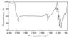 |
Fig. 4 IR spectrum of compound S1 |
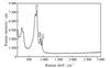 |
Fig. 5 Raman spectrum of compound S1 |
The UV/Vis of compound S1 in aqueous solution showed a high-intensity band in the visible region, centered approximately at 435 nm (Fig. 6), as has been observed in other RuIII polyoxoanions[33-36]. To identify the W/Ru oxidation states, XPS was employed. In the XPS spectra of compound S1, W 4f region displayed two partially overlapped peaks positioned at 35.18 and 37.38 eV, which are assigned to 4f7/2 and 4f5/2 of the W(VI) center (Fig. 7(a))[37]. Moreover, the binding energy peaks at 282.38 and 286.48 eV corresponds to 3d5/2 and 3d3/2 of the Ru(III) center, respectively (Fig. 7(b))[31].
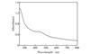 |
Fig. 6 UV/Vis absorption spectrum of aqueous solution of compound S1 |
 |
Fig. 7 XPS spectra of W 4f (a) and Ru 3d (b) in compound S1 |
2.2 Luminescence Spectra Studies
The solution-state fluorescent of S1 was investigated by fluorescence spectrum. Upon excitation at its excitation maxima, compound S1 exhibited an emission band at 685 nm. The emission profile and emission maxima were similar and independent of the excitation wavelength. The electronic emission spectral of compound S1 was presented in Fig. 8.
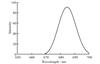 |
Fig. 8 Fluorescence emission spectrum of the S1 in solution |
2.3 Cyclic Voltammetry
To obtain the spectroscopic data, the redox behavior of compound S1 was investigated in a mixed electrolyte solution (pH = 3.0) containing Na2SO4 (0.5 mol/L) and H2SO4 (1 mol/L). Results showed that the cyclic voltammogram of RuCl3·nH2O did not exhibit any redox peak from -0.5 to 1.0 V. Subsequently, cyclic voltammetry (CV) measurements of compound S1 were conducted at a scan rate of 50 mV/s within the potential range from +1 000 to -700 mV in sulfate buffers with pH = 3 (Fig. 9). As shown in Fig. 9, the mean peak potentials (E1/2 = (Ecp + Eap)/2) for the I-I' reversible redox peaks are 300 mV, which was assignable to the RuIII/II couple[38].
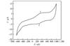 |
Fig. 9 Cyclic voltammograms of compound S1 |
2.4 Cytotoxicity Assay in vitro
The cytotoxicity of the S1, S2 (β-Na9HSiW9O34·12H2O) and S3 (RuCl3·nH2O) was determined in C33A, DLD-1, HepG2 and MRC-5 cell lines by MTT cell survival assay. These cells were treated with different concentrations of es S1, S2 and S3 for 24 h, 48 h and 72 h. As shown in Fig. 10(a), the es showed weak activities to the three cell lines for 24 h. The IC50 values of S1 were 74.06 µmol/L for C33A, 89.05 µmol/L for DLD-1 and more than 100 µmol/L for HepG2. The IC50 values of S2 and S3 to three cell lines were more than 100 µmol/L. Over a period of 48 h (as shown in Fig. 10(b)), compound S1 exhibited relatively weak activity against the C33A and DLD-1 cell lines, with IC50 values of 75.72 µmol/L and 89.73 µmol/L, respectively. Similarly, both S2 and S3 showed the weak activity to the three cell lines, with IC50 values close to 100 µmol/L. Upon extending the treatment to 72 h (Fig. 10(c)), the IC50 of the S1 was 45.48 μmol/L for C33A, 78.32 μmol/L for DLD-1 and 95.08 μmol/L for HepG2. Notably, none of complexes have cytotoxicity to the normal cell MRC-5 in different time points. The IC50 values of S1, S2 and S3 to these cells were displayed in Table 1. Cytotoxicity of some Keggin and lacunary-Keggin sandwiched polyoxotungstates towards Madin-Darby canine kidney (MDCK), Vero, HepG2, and MT-4 cells was investigated by using MTT. Results showed that none of the compounds exhibited toxicity at concentrations below 200 µmol/L in MDCK, Vero, and HepG2 cells[39]. These analyses exhibited that the complexes we studied possessed greater antitumor activity than previously reported. Obviously, the cell viability was both concentration-dependent and time-dependent, which indicated that compound S1 entered the cells slowly exerted a gradual killing effect. During the three cell lines, S1 showed the highest activity against C33A, followed by DLD-1 and HepG2. The differential cytotoxicity of these compounds toward the C33A, DLD-1 and HepG2 cell lines may be caused by their structural differences. The in vitro activity of anticancer drugs is often partially associated with their lipophilic character. And the resulting hydrophobicity could increase cellular uptake of the complex, thereby enhancing the antiproliferative activity. Alternatively, owing to the higher negative mitochondrial membrane potential present in cancer cells, lipophilic cations may preferentially cross the membrane and accumulate in mitochondria, leading to the observed higher uptake in cancer cells.
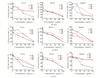 |
Fig. 10 Cell viability inhibition induced by S1, S2 and S3 C33A, DLD-1 and HepG2 cells were treated with S1, S2 and S3 for 24 h (a), 48 h (b) and 72 h (c). Data from three independent experiments |
IC50values of compounds against human tumor cell lines and MRC-5 (unit:μmol/L)
2.5 Apoptosis Studies by Hoechst 33342 Staining and Flow Cytometry
Hoechst 33342, which stains the cell nucleus, is a membrane permeable dye with blue fluorescence. Under fluorescence microscopy, live cells exhibit uniformly light blue nuclei after Hoechst 33342 treatment, while apoptotic cells display bright blue nuclei due to karyopyknosis and chromatin condensation. Conversely, dead cells' nuclei remain unstained[40]. Subsequently, C33A cells were treated with S1 at 5 µmol/L, 25 µmol/L and 50 µmol/L for 48 h and followed by stained with Hoechst 33342. As shown in Fig. 11, under control conditions, cells displayed homogeneous nuclear staining. In contrast, treatment with S1 resulted in a dose-dependent increase in apoptotic C33A cells, characterized by bright staining, condensed chromatin, and fragmented nuclei. These results show that compound S1 effectively induces apoptosis in C33A cells.
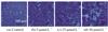 |
Fig.11 Hoechst 33342 staining of C33A cells induced by S1 |
To determine the percentages of apoptotic and necrotic cells, untreated C33A cells served as control, and apoptosis was investigated by flow cytometry (as shown in Fig. 12). In the control, the proportions of living cells and apoptotic cells were 99.6% and 0.3%, respectively. When C33A cells were exposed to compound S1 (5, 25, 50 µmol/L) for 48 h, the proportions of apoptotic cells were 3.6%, 11.6% and 54.5%, respectively. Compared with the control, the proportion of living cells decreased and apoptotic cells increased. These data demonstrate that the apoptotic effect of compound S1 on C33A cells exhibits a positive dose-responsive manner.
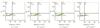 |
Fig. 12 The percentage of living (C3), late apoptotic (C2), and early apoptotic (C4) in C33A cells |
2.6 Cell Cycle Arrest
Flow cytometry analysis was utilized to assess the distribution of C33A cells in different phases of the cell cycle followed by propidium iodide staining. As shown in Fig. 13, treatment of C33A cells with 5 µmol/L, 25 µmol/L, and 50 µmol/L of compound S1 for 48 h resulted in a significant augmentation in the percentage of cells in the S phase, with increases of 2.5%, 7.9% and 16.5%, respectively. Moreover, compared to the control, treatment of C33A cells with 50 µmol/L of compound S1 for 48 h led to a decrease of 20.5% in the G0/G1 phase. These data demonstrate that S1 induce S phase arrest in C33A cells[41].
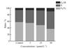 |
Fig. 13 Cell cycle distribution of C33A cells induced by S1 |
3 Conclusion
In summary, the introduction of ruthenium into trilacunary POMs reaction system generated a ruthenium multi-substituted polyoxometalates. In vitro, compound S1 exhibits higher cytotoxicity toward C33A cell line compared to DLD-1 and HepG2 cell lines. Hoechst 33342 staining demonstrates that compound S1 effectively induces apoptosis in C33A cells. Apoptosis assay demonstrate a concentration-dependent increase in apoptotic cells upon treatment with compound S1. Furthermore, cell cycle arrest studies reveal that the antiproliferative effect of compound S1 on C33A cells occured in S phase. These findings suggest that ruthenium-substituted polyoxometalates can be used as new potential candidate for chemotherapy drugs in tumor therapy. However, the underlying mechanism of compound S1 on apoptotic induction in cancer cells needs further investigation.
References
- Huang X L, Zhang Z H, Jia L, et al. Endoplasmic reticulum stress contributes to vitamin E succinate-induced apoptosis in human gastric cancer SGC-7901 cells[J]. Cancer Letters, 2010, 296(1): 123-131. [CrossRef] [PubMed] [Google Scholar]
- Liu H K, Wang Q, Li Y, et al. Inhibitory effects of γ-tocotrienol on invasion and metastasis of human gastric adenocarcinoma SGC-7901 cells[J]. The Journal of Nutritional Biochemistry, 2010, 21(3): 206-213. [CrossRef] [PubMed] [Google Scholar]
- Ghobrial I M, Witzig T E, Adjei A A. Targeting apoptosis pathways in cancer therapy[J]. CA: A Cancer Journal for Clinicians, 2005, 55(3): 178-194. [CrossRef] [PubMed] [Google Scholar]
- Hasenknopf B. Polyoxometalates: Introduction to a class of inorganic compounds and their biomedical applications[J]. Frontiers in Bioscience: A Journal and Virtual Library, 2005, 10: 275-287. [CrossRef] [Google Scholar]
- Čolović M B, Lacković M, Lalatović J, et al. Polyoxometalates in biomedicine: Update and overview[J]. Current Medicinal Chemistry, 2020, 27(3): 362-379. [CrossRef] [PubMed] [Google Scholar]
- Gerth H U V, Rompel A, Krebs B, et al. Cytotoxic effects of novel polyoxotungstates and a platinum compound on human cancer cell lines[J]. Anti-Cancer Drugs, 2005, 16(1): 101-106. [CrossRef] [PubMed] [Google Scholar]
- Wang J, Liu Y, Xu K, et al. Broad-spectrum antiviral property of polyoxometalate localized on a cell surface[J]. ACS Applied Materials & Interfaces, 2014, 6(12): 9785-9789. [CrossRef] [PubMed] [Google Scholar]
- Herve M, Sinoussi-Barre F, Chermann J C, et al. Correlation between structure of polyoxotungstates and their inhibitory activity on polymerases[J]. Biochemical and Biophysical Research Communications, 1983, 116(1): 222-229. [CrossRef] [PubMed] [Google Scholar]
- Aureliano M, Gândara R M C. Decavanadate effects in biological systems[J]. Journal of Inorganic Biochemistry, 2005, 99(5): 979-985. [CrossRef] [PubMed] [Google Scholar]
- Lee I S, Long J R, Prusiner S B, et al. Selective precipitation of prions by polyoxometalate complexes[J]. Journal of the American Chemical Society, 2005, 127(40): 13802-13803. [NASA ADS] [CrossRef] [PubMed] [Google Scholar]
- Müller C E, Iqbal J, Baqi Y, et al. Polyoxometalates—A new class of potent ecto-nucleoside triphosphate diphosphohydrolase (NTPDase) inhibitors[J]. Bioorganic & Medicinal Chemistry Letters, 2006, 16(23): 5943-5947. [CrossRef] [PubMed] [Google Scholar]
- Balula S S, Santos I C M S, Barbosa A D S, et al. Manganese mono-substituted borotungstate: Characterization and catalytic application[J]. Materials Science Forum, 2012, 730: 975-980. [CrossRef] [Google Scholar]
- Kikukawa Y, Suzuki K, Yamaguchi K, et al. Synthesis, structure characterization, and reversible transformation of a cobalt salt of a dilacunary γ-keggin silicotungstate and sandwich-type di- and tetracobalt-containing silicotungstate dimers[J]. Inorganic Chemistry, 2013, 52(15): 8644-8652. [CrossRef] [PubMed] [Google Scholar]
- Dutta D, Jana A D, Debnath M, et al. Design of tri-substituted dodecatungstosilicate from a trilacunary silicotungstate by insertion of manganese ions of [Mn3(μ3-O)(2-Cl-benzoato)6(py)3]: Synthesis, structure, redox and magnetic studies[J]. European Journal of Inorganic Chemistry, 2010, 35: 5517-5522. [CrossRef] [Google Scholar]
- Wang L, Zhou B B, Yu K, et al. Novel antitumor agent, trilacunary Keggin-type tungstobismuthate, inhibits proliferation and induces apoptosis in human gastric cancer SGC-7901 cells[J]. Inorganic Chemistry, 2013, 52(9): 5119-5127. [CrossRef] [PubMed] [Google Scholar]
- Wang L, Yu K, Zhou B B, et al. The inhibitory effects of a new cobalt-based polyoxometalate on the growth of human cancer cells[J]. Dalton Transactions, 2014, 43(16): 6070-6078. [CrossRef] [PubMed] [Google Scholar]
- Sava G, Bergamo A. Ruthenium-based compounds and tumour growth control (review)[J]. International Journal of Oncology, 2000, 17(2): 353-365. [PubMed] [Google Scholar]
- Rademaker-Lakhai J M, van den Bongard D, Pluim D, et al. A Phase I and pharmacological study with imidazolium-trans-DMSO-imidazole-tetrachlororuthenate, a novel ruthenium anticancer agent[J]. Clinical Cancer Research: An Official Journal of the American Association for Cancer Research, 2004, 10(11): 3717-3727. [CrossRef] [PubMed] [Google Scholar]
- Sadakane M, Tsukuma D, Dickman M H, et al. Dimerization of mono-ruthenium substituted α-Keggin-type tungstosilicate[α-SiW11O39RuIII(H2O)]5- to µ-oxo-bridged dimer in aqueous solution: Synthesis, structure, and redox studies[J]. Dalton Transactions, 2007, 26: 2833-2838. [CrossRef] [Google Scholar]
- Yokoyama A, Ohkubo K, Ishizuka T, et al. Remarkable enhancement of catalytic activity of a 2∶1 complex between a non-planar Mo(v)-porphyrin and a ruthenium-substituted Keggin-type heteropolyoxometalate in catalytic oxidation of benzyl alcohols[J]. Dalton Transactions, 2012, 41(33): 10006-10013. [CrossRef] [PubMed] [Google Scholar]
- Lahootun V, Besson C, Villanneau R, et al. Synthesis and characterization of the keggin-type ruthenium-nitrido derivative [PW11O39{RuN}]4- and evidence of its electrophilic reactivity[J]. Journal of the American Chemical Society, 2007, 129(22): 7127-7135. [NASA ADS] [CrossRef] [PubMed] [Google Scholar]
- Liu B, Yan J, Wang Y F, et al. Redox chemistry of ruthenium ions in mono-substituted Keggin tungstophosphate: A new synthetic extension for ruthenium derivatives based on[PW11O39Ru(VI)N](4.)[J]. Dalton Transactions, 2015, 44(38): 16882-16887. [CrossRef] [PubMed] [Google Scholar]
- Liu H Z, Yue B, Sun W L, et al. Synthesis and characterization of noble-metal-substituted Dawson-type polyoxometalates[J]. Transition Metal Chemistry, 1997, 22(4): 321-325. [CrossRef] [Google Scholar]
- Nomiya K, Torii H, Nomura K, et al. Synthesis and characterization of a monoruthenium(III)-substituted Dawson polyoxotungstate derived by Br2 oxidation of the 1∶2 complex of ruthenium(II) and [α2-P2W17O61]10-. The reactivity of cis-[RuCl2(DMSO)4] as a ruthenium source[J]. Journal of the Chemical Society, Dalton Transactions, 2001, 9: 1506-1512. [CrossRef] [Google Scholar]
- Ogo S, Shimizu N, Ozeki T, et al. Determination of α-Keggin structure of [GeW11O39RuIII(H2O)]5-. Reaction of[GeW11O39RuIII(H2O)]5- with dimethyl sulfoxide to form[GeW11O39RuIII(dmso)]5- and their structural characterization[J]. Dalton Trans, 2013, 42(7): 2540-2545. [CrossRef] [PubMed] [Google Scholar]
- Howells A R, Sankarraj A, Shannon C. A diruthenium-substituted polyoxometalate as an electrocatalyst for oxygen generation[J]. Journal of the American Chemical Society, 2004, 126(39): 12258-12259. [NASA ADS] [CrossRef] [PubMed] [Google Scholar]
- Morris A M, Anderson O P, Finke R G. Reinvestigation of a Ru2-incorporated polyoxometalate dioxygenase precatalyst, "[WZnRu2III(H2O)(OH)(ZnW9O34)2]11-": Evidence for marginal, ≤ 0.2 equivalents of Ru incorporation plus faster catalysis by physical mixtures of [RuII(DMSO)4Cl2] and the parent polyoxometalate [WZn3(H2O)2(ZnW9O34)2]12-[J]. Inorganic Chemistry, 2009, 48(10): 4411-4420. [CrossRef] [PubMed] [Google Scholar]
- Jia S F, Hao X L, Wen Y Z, et al. Synthesis, cytotoxicity, apoptosis and cell cycle arrest of a monoruthenium(II)-substituted Dawson polyoxotungstate[J]. Journal of Coordination Chemistry, 2019, 72(4): 633-644. [CrossRef] [MathSciNet] [Google Scholar]
- Tézé A, Hervé G. α-, β-, and γ-dodecatungstosilicic acids: isomers and related lacunary compounds[J]. Inorganic Syntheses, 1990, 27: 88-91. [Google Scholar]
- Mosmann T. Rapid colorimetric assay for cellular growth and survival: Application to proliferation and cytotoxicity assays[J]. Journal of Immunological Methods, 1983, 65: 55-63. [CrossRef] [PubMed] [Google Scholar]
- Gamelas J A F, Carapuça H M, Balula M S, et al. Synthesis and characterisation of novel ruthenium multi-substituted polyoxometalates: α, β-[SiW9O37Ru4(H2O)3Cl3]7-[J]. Polyhedron, 2010, 29(16): 3066-3073. [CrossRef] [Google Scholar]
- Kortz U, Tézé A, Hervé G. A cubane-substituted polyoxoanion: Structure and magnetic properties of Cs2[H2PW9Ni4O34(OH)3(H2O)6]·5H2O[J]. Inorganic Chemistry, 1999, 38(9): 2038-2042. [CrossRef] [PubMed] [Google Scholar]
- Neumann R, Abu-Gnim C. Alkene oxidation catalyzed by a ruthenium-substituted heteropolyanion, SiRu(L)W11O39: The mechanism of the periodate-mediated oxidative cleavage[J]. Journal of the American Chemical Society, 1990, 112(16): 6025-6031. [NASA ADS] [CrossRef] [Google Scholar]
- Sadakane M, Higashijima M. Synthesis and electrochemical behavior of [SiW11O39RuIII(H2O)]5- and its oxo-bridged dimeric complex [SiW11O39RuIVORuIIISiW11O39]11-[J]. Dalton Transactions, 2003, 4: 659-664. [CrossRef] [Google Scholar]
- Neumann R, Khenkin A M, Dahan M. Hydroxylation of alkanes with molecular oxygen catalyzed by a new ruthenium-substituted polyoxometalate, [WZnRu(OH)(H2O)(ZnW9O34)2]11-[J]. Angewandte Chemie International Edition, 1995, 34(15): 1587-1589. [NASA ADS] [CrossRef] [Google Scholar]
- Quiñonero D, Wang Y, Morokuma K, et al. The role of the central atom in structure and reactivity of polyoxometalates with adjacent d-electron metal sites. computational and experimental studies of γ-[(Xn+O4)RuIII2(OH)2(MFM)10O32](8-n)- for MFM = Mo and W, and X = AlIII, SiIV, PV, and SVI[J]. The Journal of Physical Chemistry B, 2006, 110(1): 170-173. [CrossRef] [PubMed] [Google Scholar]
- Hao X L, Ma Y Y, Wang Y H, et al. New entangled coordination networks based on charge-tunable keggin-type polyoxometalates[J]. Chemistry—An Asian Journal, 2014, 9(3): 819-829. [CrossRef] [PubMed] [Google Scholar]
- Landsmann S, Wessig M, Schmid M, et al. Smart inorganic surfactants: More than surface tension[J]. Angewandte Chemie International Edition, 2012, 51(24): 5995-5999. [CrossRef] [PubMed] [Google Scholar]
- Yamase T. Anti-tumor, -viral, and-bacterial activities of polyoxometalates for realizing an inorganic drug[J]. Journal of Materials Chemistry, 2005, 15(45): 4773. [CrossRef] [Google Scholar]
- Li J F, Huang R Z, Yao G Y, et al. Synthesis and biological evaluation of novel aniline-derived Asiatic acid derivatives as potential anticancer agents[J]. European Journal of Medicinal Chemistry, 2014, 86: 175-188. [CrossRef] [PubMed] [Google Scholar]
- Zhang Y, Hu P C, Cai P, et al. Synthesis, characterization, crystal structure, cytotoxicity, apoptosis and cell cycle arrest of ruthenium(ii) complex [Ru(bpy)2(adpa)](PF6)2 (bpy = 2, 2'-bipyridine, adpa = 4-(4-aminophenyl)diazenyl-N-(pyridin-2-ylmethylene)aniline)[J]. RSC Advances, 2015, 5(15): 11591-11598. [NASA ADS] [CrossRef] [Google Scholar]
All Tables
All Figures
|
Fig. 1 SEM image (a) and EDS (b) of compound S1 |
| In the text | |
|
Fig. 2 TG curve of compound S1 |
| In the text | |
|
Fig. 3 Possible structure of the Ru tetra-substituted anions [SiW9O37Ru4(H2O)3Cl3]7- in compound S1 |
| In the text | |
|
Fig. 4 IR spectrum of compound S1 |
| In the text | |
|
Fig. 5 Raman spectrum of compound S1 |
| In the text | |
|
Fig. 6 UV/Vis absorption spectrum of aqueous solution of compound S1 |
| In the text | |
|
Fig. 7 XPS spectra of W 4f (a) and Ru 3d (b) in compound S1 |
| In the text | |
|
Fig. 8 Fluorescence emission spectrum of the S1 in solution |
| In the text | |
|
Fig. 9 Cyclic voltammograms of compound S1 |
| In the text | |
|
Fig. 10 Cell viability inhibition induced by S1, S2 and S3 C33A, DLD-1 and HepG2 cells were treated with S1, S2 and S3 for 24 h (a), 48 h (b) and 72 h (c). Data from three independent experiments |
| In the text | |
|
Fig.11 Hoechst 33342 staining of C33A cells induced by S1 |
| In the text | |
|
Fig. 12 The percentage of living (C3), late apoptotic (C2), and early apoptotic (C4) in C33A cells |
| In the text | |
|
Fig. 13 Cell cycle distribution of C33A cells induced by S1 |
| In the text | |
Current usage metrics show cumulative count of Article Views (full-text article views including HTML views, PDF and ePub downloads, according to the available data) and Abstracts Views on Vision4Press platform.
Data correspond to usage on the plateform after 2015. The current usage metrics is available 48-96 hours after online publication and is updated daily on week days.
Initial download of the metrics may take a while.